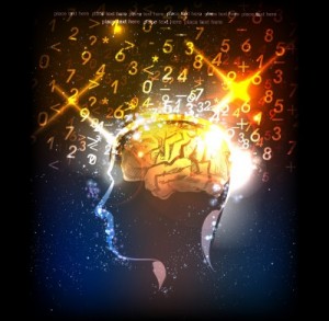
 Brain Mapping is trying to understand which parts of the brain are active and which are not so active. We need to understand the anatomy of the brain to exactly get to the bottom of brain mapping.
The spinal cord and the brain make up for the central nervous system, which regulates both our body and mind. The central nervous system helps us coordinate our limbs and all that we do in our day to day lives. The Brain Mapping helps us to understand what exactly goes in ones mind. It also helps us to interpret information from the environment and is the basis of the thought process and our emotional being. Each part of the brain, its functions, understanding the fluids that support and provide nourishment, exploring its anatomy, is all a part of the brain mapping.
One can avail the diagrammatic expression of what brain mapping is all about. Brain anatomy helps you to get an insight into the concept which is so unique and fascinating. The diagrams are so interactive that they provide a flexible visual image so that the concepts can be well communicated to parents, students and children. A human brain has a knot of 100 billion neurons and support cells, which help us to cherish our memories there. This is what helps us to be creative and thus write sonnets or mastermind big projects like building planes. An elephant’s brain is larger, weighing more than a human brain, and has more neurons than us, but they are unable to perform all that we can perform. This is the prime reason that brain mapping of a human brain is becoming so important.
Brain Mapping relates the brain structure to its function and to understand which part helps us to do what. Which part of our brain helps us to reason, logic or be creative. This can be termed as localization of function. One part of the brain helps us to listen and the other helps us to look, observe and see. A question may arise as to which part of the brain helps us to distinguish between colors. Brain mapping also looks into the brain from outside, the changes that take place due to environmental factors like learning, aging. It also helps you to examine the portions that are being affected during a mental illness or any other brain related disease.
Brain Mapping gives us a thorough picture of the brain, its structure and its functional lobes, neuron bundles which connect brain parts, neuron circuits junctions between the neurons and the parts of the neurons. The brain mapping is a collection of many tools. The researchers collect images, turn these images into data, and then analyze this data to see what happens in the brain.
Brain Mapping is a set of neuroscience techniques which are predicted on the mapping of biological quantities onto representations, which results in maps. This can be further defined as a study of anatomy and the functions of spinal cord and the brain through the use of imaging. Neuro imaging MRI are all a part of brain mapping.
The methodologies are constantly being invented and reinvented. Functions and neuroimaging form the base of Brain mapping. This is being used by the doctors to plan surgeries which are safe. Epilepsy is being treated effectively by removing the affected parts of the brain through the Brain Mapping. MRI and EEG helps the surgeons to locate seizure center in a patient’s brain, as well as the areas that are active during speaking and moving. The images help the doctor to chalk out exactly what to remove and what to keep. This also helps in diagnosing neurodegenerative diseases like Alzheimer’s and Parkinson’s, using techniques like PET, MRI to understand the shrinkages which show tissue loss. Autism too can be understood clearly. Diagrams can be drawn using this technique to find out how, when and the extent of autism.
The scientists use many ways to study the structure of the brain and the functions. They compare the picture of a healthy brain with that of a diseased brain. They also examine neurons at the microscopic level.
The tools that are used can be a) Computer axial tomography (CAT)-these are X-rays of the brain from various angles to understand structural abnormalities b) Structural magnetic resonance imaging which takes advantage of the fluids in the brain to create images which have a better resolution than a CAT scan c) Diffusion tensor – MRI (DTI) images of neurons and its tracts.